
3D X-Ray Diagnostics in Lucerne
For implants, complex root canal work and oral surgery, precise 3D imaging is the best foundation. At our practice in Lucerne we use modern CBCT devices with significantly reduced radiation dose.
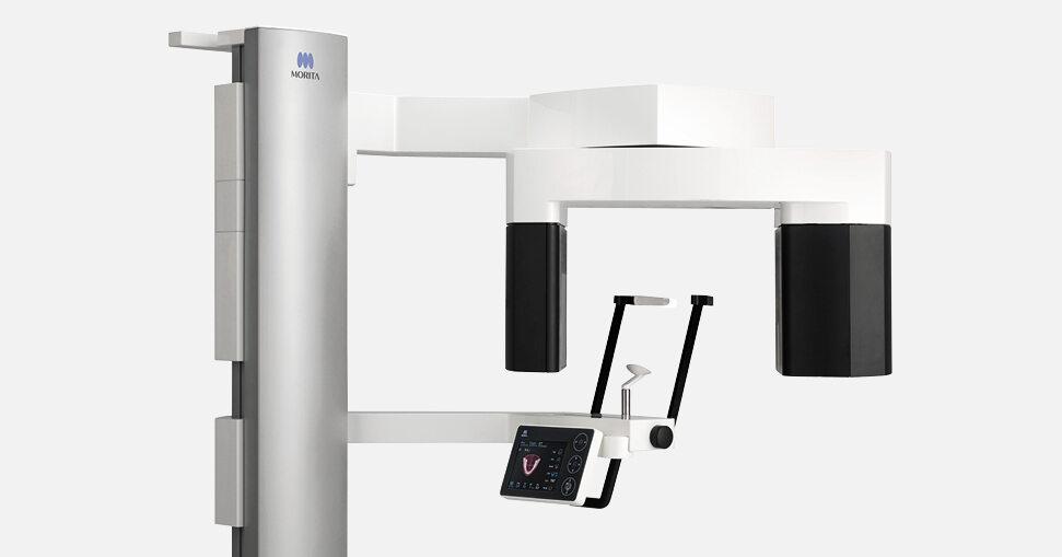
3D X-Ray Diagnostics (CBCT) in Lucerne
3D scans in Lucerne allow safe planning, reduce surgical risks and shorten treatment times – for more safety and fewer surprises.
- Three-dimensional imaging of bone, teeth and nerves
- Low-dose CBCT with state-of-the-art sensor technology
- Ideal for implant planning, endodontics and wisdom teeth
- Digital planning directly on screen
What is 3D X-ray diagnostics?
3D X-ray diagnostics, also known as cone beam computed tomography (CBCT), offers several advantages over conventional 2D X-ray procedures. Instead of a flat image, it produces a three-dimensional data model that can be viewed from all directions and slice by slice.
In dentistry the procedure is usually called digital volume tomography (DVT). What happens during a scan, what it shows and how high the radiation exposure is are explained in detail under digital volume tomography (DVT).
Advantages over 2D X-rays
Enhanced spatial imaging
3D X-ray images allow a more detailed and more accurate spatial representation of structures than conventional 2D X-rays.
More precise diagnosis
The three-dimensional representation allows a much more precise assessment of the anatomical situation. The data can be measured 1:1 and processed in planning software.
Lower radiation exposure
Although 3D X-rays deliver more information, the radiation exposure can often be lower because the imaging is more efficient and targeted.
Better planning of procedures
Precise 3D images allow more accurate advance planning of surgical procedures and other treatments. This prevents unpleasant surprises during a procedure.
2D and 3D X-rays compared
| Feature | 2D X-ray | 3D X-ray (DVT) |
|---|---|---|
| Image | Flat, two-dimensional image | Spatial, three-dimensional model |
| Overlapping | Structures can overlap | Slices can be viewed individually |
| Measurement | Only to a limited extent | Measurable 1:1, usable in planning software |
| Typical use | Overview images, checking individual teeth | Implant planning, wisdom teeth, root canals, surgical procedures |
What we use 3D X-rays for
- Implantology: planning the position, length and angle of a dental implant, combined with a drilling guide if required (guided surgery).
- Endodontics: assessing root canals and inflammation at the root tip, for example before microscopic root canal treatment.
- Oral surgery: the position of wisdom teeth and nerves before a procedure, for example in microsurgery.
- Maxillofacial surgery: assessing the jawbone and maxillary sinus as well as cysts and bone defects.
- Digital planning: together with the scan from impression-free dentistry, it creates a complete digital model for treatment planning.
High image sharpness with the Veraview X800
The sharper the images, the more precise the diagnosis – and the more accurately treatment can be planned.
We work with the Veraview X800 dental X-ray system. It achieves a high resolution (> 2.5 LP/mm) with a voxel size of 80 μm and provides a reliable basis for diagnosis in all areas of dentistry, from implantology and endodontics to oral and maxillofacial surgery.
Frequently asked questions about 3D X-rays
Answers on uses, radiation exposure, procedure and cost of 3D X-ray diagnostics.
What is the difference between a 3D X-ray and DVT?
In dentistry both terms usually mean the same thing: digital volume tomography (DVT), internationally called cone beam computed tomography (CBCT), is the 3D X-ray procedure for teeth and jaw. Detailed information is available under digital volume tomography.
When is a 3D X-ray useful?
A 3D X-ray is useful when a 2D image is not enough for diagnosis or planning – for example before an implant, with impacted wisdom teeth, complex root canal treatment or suspected cysts and bone defects. We discuss with you in advance whether a scan is needed in your case.
Are dental 3D X-rays harmful?
DVT works with comparatively low radiation exposure, often lower than a conventional CT. We only take a 3D scan when it brings a benefit for diagnosis or planning. In pregnant women it is only used in urgent cases.
What happens during a 3D X-ray?
You stand or sit at the unit, with your chin and forehead supported. During the scan the unit rotates once around your head; the scan is painless. Software then combines the individual images into a 3D model, which we discuss with you.
How much does a dental 3D X-ray cost?
The cost depends on the extent of the scan and its evaluation. If the scan is part of a treatment, such as an implant, it is listed in the cost estimate. Basic health insurance does not usually cover dental treatment; supplementary dental insurance may contribute, depending on your policy.
Where does LifeDent offer 3D X-ray diagnostics?
At our practices in Lucerne and Volketswil. You can request an appointment online for an examination.


